We are a research group located at the Center for MR-Research, University Children's Hospital Zürich / University of Zürich. We are interested in how novel technologies, such as in vivo fetal MRI, can support clinical decision making and how the in vivo – in utero approach can extend our current knowledge of human brain development.
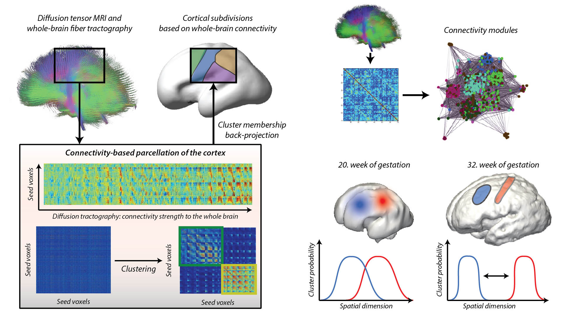
Connectivity architecture of the developing human brain
Fetal magnetic resonance imaging (MRI) is a cutting-edge, noninvasive medical diagnostic procedure that is used to diagnose abnormalities before birth. Recent advancements in imaging technology enabled the adaptation of the so-called diffusion-MRI approach to developing fetuses. Our team has successfully used diffusion tensor MRI (DT-MRI) for fetuses during clinical practice and develops the necessary image analysis and image processing software. This method allows us to visualize the 3D structure of the developing fiber connectivity of the fetal brain in pathologies but also in normally developing fetal brains.
Jakab A, Kasprian G, Schwartz E, Gruber GM, Mitter C, Prayer D, Schöpf V, Langs G. Disrupted developmental organization of the structural connectome in fetuses with corpus callosum agenesis. Neuroimage. 2015 May 1;111:277-88.
Jakab A, Pogledic I, Schwartz E, Gruber G, Mitter C, Brugger PC, Langs G, Schöpf V, Kasprian G, Prayer D. Fetal Cerebral Magnetic Resonance Imaging Beyond Morphology. Semin Ultrasound CT MR. 2015 Dec;36(6):465-75.
Jakab A, Schwartz E, Kasprian G, Gruber GM, Prayer D, Schöpf V, Langs G. Fetal functional imaging portrays heterogeneous development of emerging human brain networks. Front Hum Neurosci. 2014 Oct 22;8:852.
Jakab A, Molnár PP, Bogner P, Béres M, Berényi EL. Connectivity-based parcellation reveals interhemispheric differences in the insula. Brain Topogr. 2012 Jul;25(3):264-71.
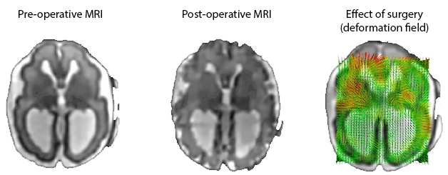
Neurodevelopment after fetal surgery
Spina bifida (myelomeningocele and myeloschisis) is a common birth defect that is associated with significant lifelong morbidity, and it has only recently become possible to perform open fetal surgical correction of the underlying anatomical abnormality to prevent the loss of neurological function. We assume that the corrective surgery, which restores the normal developmental milieau and the hydrodynamic pressure of the ventricular system, has an impact on the development of sensory-motor systems of the brain as well. A central hypothesis is that in addition to the purely morphological effect of the corrective surgery, such as the normalisation of cerebrospinal fluid space volumes, neuronal maturation, formation of synapses, and the overall brain development are also affected. This may cause detectable changes in tissue microarchitecture (such as diffusivity), brain fiber anatomy and brain connectivity.
Rethmann C, Scheer I, Meuli M, Mazzone L, Moehrlen U, Kellenberger CJ. Evolution of posterior fossa and brain morphology after in utero repair of open neural tube defects assessed by MRI. Eur Radiol. 2017 Nov;27(11):4571-4580.
Meuli M, Moehrlen U. Fetal surgery for myelomeningocele is effective: a critical look at the whys. Pediatr Surg Int. 2014 Jul;30(7):689-97.
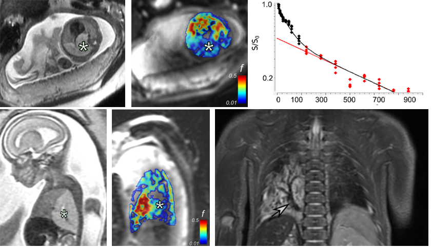
Functional imaging of the fetal body, brain and placenta
We implemented diffusion-weighted and intravoxel incoherent motion MRI sequences to capture how the function (e.g. perfusion) of the placenta and fetal organs change during gestation. We aim to investigate if such quantitative markers are affected by common conditions, such as intrauterine growth retardation or congenital disorders affecting brain maturation.
Jakab A, Tuura R, Kottke R, Kellenberger CJ, Scheer I. Intra-voxel incoherent motion MRI of the living human foetus: technique and test-retest repeatability. Eur Radiol Exp. 2017;1(1):26.
Jakab A, Tuura RL, Kottke R, Ochsenbein-Kölble N, Natalucci G, Nguyen TD, Kellenberger C, Scheer I. Microvascular perfusion of the placenta, developing fetal liver, and lungs assessed with intravoxel incoherent motion imaging. J Magn Reson Imaging. 2018 Jul;48(1):214-225.
Semester and master thesis topics
We offer thesis supervision (ETH and or University of Zürich) in the following clinical and basic research topics:
- Development of the fetal brain connectome (background: MD, biology)
- Effect of open fetal surgery on brain morphology (background: MD, biology)
- Motion correction for fetal diffusion tensor imaging (background: engineering)
If you are interested, feel free to contact us!